Home

rolka Cordelia uczestniczyć color fundus optos צילום צבע Luminancja spotykać się odebrać

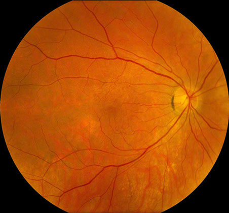
Color Fundus | Optique of Denver

July-2010
July-2010

Optos color photographs OD and OS (above), demonstrating vascular... |  Download Scientific Diagram
Optos color photographs OD and OS (above), demonstrating vascular... | Download Scientific Diagram

Optos color photographs OD and OS (above), demonstrating vascular... |  Download Scientific Diagram
Optos color photographs OD and OS (above), demonstrating vascular... | Download Scientific Diagram

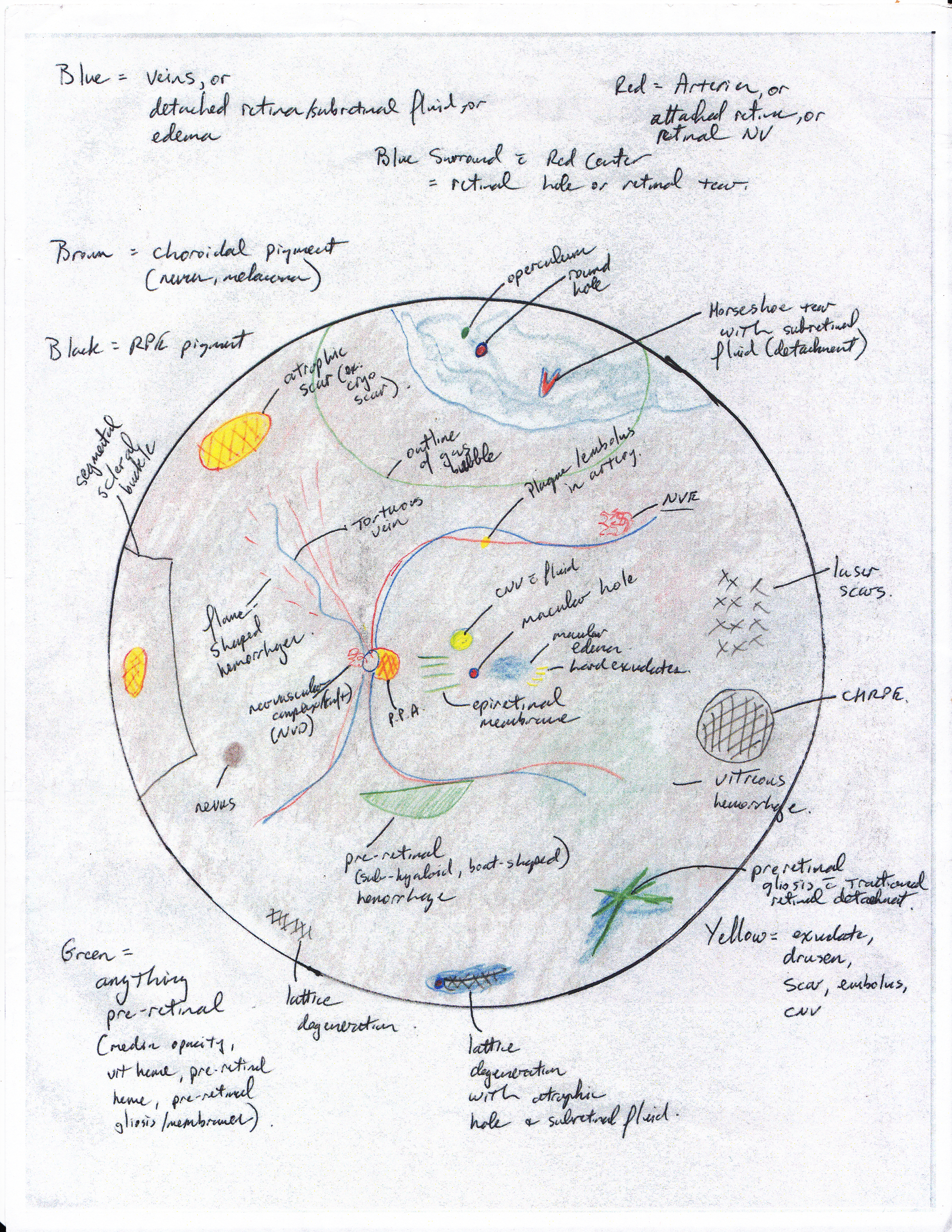
Basics of Fundus (Retina) Drawing | ShortWhiteCoats.com

Optos color photographs OD and OS (above), demonstrating vascular... |  Download Scientific Diagram
Optos color photographs OD and OS (above), demonstrating vascular... | Download Scientific Diagram

Examples of the variation of color among fundus images of the same retina.  | Download Scientific Diagram
Examples of the variation of color among fundus images of the same retina. | Download Scientific Diagram

Diagnostic Atlas af Booklet - A Reference Guide - Flip eBook Pages 1-36 |  AnyFlip
Diagnostic Atlas af Booklet - A Reference Guide - Flip eBook Pages 1-36 | AnyFlip

Pseudo-colour fundal Optos images of a baby with stage 3, zone-2 ROP... |  Download Scientific Diagram
Pseudo-colour fundal Optos images of a baby with stage 3, zone-2 ROP... | Download Scientific Diagram

Optos color photographs OD and OS (above), demonstrating vascular... |  Download Scientific Diagram
Optos color photographs OD and OS (above), demonstrating vascular... | Download Scientific Diagram

Using Retinal Imaging to Study Dementia | Protocol (Translated to Hebrew)

סריקת אופטלמוסקופיית לייזר - Scene in San Francisco - Wikipedia

Color fundus picture of the OD (a) and OS (b) showing peripapillary... |  Download Scientific Diagram
Color fundus picture of the OD (a) and OS (b) showing peripapillary... | Download Scientific Diagram

Optos color photographs OD and OS (above), demonstrating vascular... |  Download Scientific Diagram
Optos color photographs OD and OS (above), demonstrating vascular... | Download Scientific Diagram

Fundus color images of the right (a) and left (b) eye showing retinal... |  Download Scientific Diagram
Fundus color images of the right (a) and left (b) eye showing retinal... | Download Scientific Diagram

Top) Color Fundus image with anatomi- | Download Scientific Diagram

Cornell Medical Instruments - Fundus Camera and Retinal Imaging Devices

Optos color photographs OD and OS (above), demonstrating vascular... |  Download Scientific Diagram
Optos color photographs OD and OS (above), demonstrating vascular... | Download Scientific Diagram

עיוורון צבעים ואנקרומה – EnChroma

Color fundus picture of the OD (a) and OS (b) showing peripapillary... |  Download Scientific Diagram
Color fundus picture of the OD (a) and OS (b) showing peripapillary... | Download Scientific Diagram

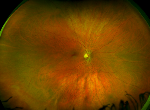
California icg
California icg

Examples of the variation of color among fundus images of the same retina.  | Download Scientific Diagram
Examples of the variation of color among fundus images of the same retina. | Download Scientific Diagram

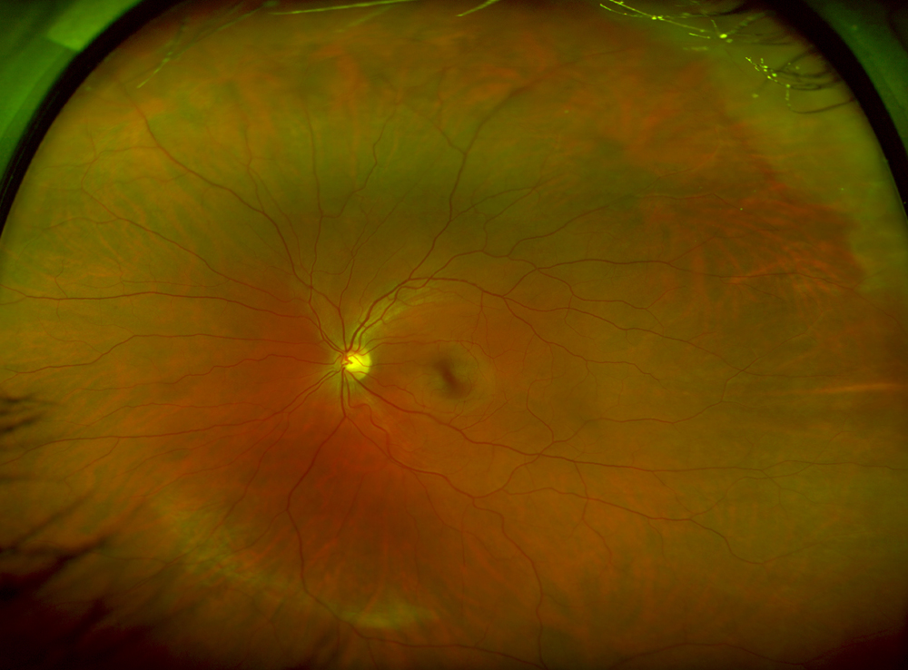
Applications of fundus autofluorescence and widefield angiography in  clinical practice - Canadian Journal of Ophthalmology
Applications of fundus autofluorescence and widefield angiography in clinical practice - Canadian Journal of Ophthalmology

Using Retinal Imaging to Study Dementia | Protocol (Translated to Hebrew)

Fluorescein angiogram of the right eye of a patient showing a... | Download  Scientific Diagram
Fluorescein angiogram of the right eye of a patient showing a... | Download Scientific Diagram

Colour fundus retinal features. | Download Scientific Diagram

California Fa - Best 200 degree Fluorescein angiography (non mydriatic FA)